| Availability: | |
|---|---|
| Quantity: | |
GEV-580
Geiwre
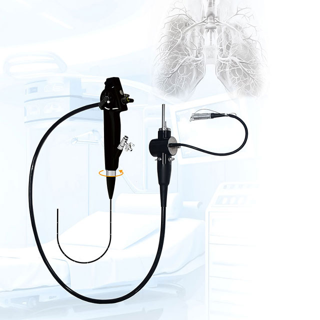
The HD Gastroscope and Colonoscope Endoscope LED Cold Light System is an advanced medical imaging device designed for comprehensive gastrointestinal examinations. Equipped with an outstanding optical system, it delivers a wide field of view and sharp, high-definition images to support accurate medical assessments. The system integrates a high-performance video processor and a reliable LED cold light source, ensuring stable illumination and efficient image processing. Featuring innovative electronic staining technology, this system enables detailed visualization of mucosal layers, facilitating the early detection and precise evaluation of potential abnormalities. This complete system provides a reliable and efficient solution for various endoscopic procedures.
Outer diameter of head end: ≤ф9.8mm / Ф7.2mm / Ф6.0mm
Outer diameter of main hose: ≤ ф9.6mm / Ф7.0mm / ф5.8mm
Inner diameter of clamp pipe: > Ф2.8mm / Ф2.4mm / ф2.4mm
Working length: ≥ 1050mm
Total length: >1335mm
Field of view: ≥ 140 °(direct view)
Observation depth: ≥ 2-100mm
Resolution: ≥ 1 million pixels CMOS HD-1080P
Bending angle: ≥ 210°upward / ≥ 100° downward / ≥ 100° left / ≥ 230° right maximum
Outer diameter of main hose: <Φ8.7mm
Outer diameter of main hose: ≤Φ12.8mm
Inner diameter of clamp pipe: 202.8mm
Inner diameter of clamp pipe: 2Φ3.75mm
Working length: 1500mm
Working length: 1350mm
Total length: ≥1650mm
Field of view: ≥160 (direct view)
Field of view: ≥170 °(direct view)
Observation depth: 22-100mm
Observation depth: 2-150mm
Resolution: ≥1,000,000 pixels
Resolution: 1,000,000 pixels
Bending angle: Up And Down≥180°
Left And Right ≥160°
Left And Right >160
Image processor: powerful, brightness, colour and contrast adjustable from 0-100 levels
Resolution: 1,000,000 pixels
Auto / Manual White balance: Auto / Manual
Video output: HDMI IN / HDMI OUT / USB
Image video storage: USB stick for direct photo and video saving
Video saving
Spectrum range: 400-700nm
LED power: 100W
LED illumination: >200,000Lx
Life: 50,000 hours
Color temperature: >5500K
Air pressure and gas flow: Air pressure: 30~60KPa, air flow: 4~10L/min
Air pump adjustment: 0 to 3 is adjustable
Power supply: AC 85~264V
LCI and NBI each represent advanced image enhancement technologies designed for precise endoscopic evaluation.
LCI is an innovative image enhancement endoscopy technology. By emphasizing slight color differences in the mucosal surface, LCI allows for quicker and more accurate identification of suspicious areas, which helps in shortening the overall procedure time. This technology offers unique advantages in observing subtle changes in mucosal morphology and color, providing critical visual support for early detection and comprehensive evaluation of gastrointestinal conditions without relying on traditional dyes.
NBI is an emerging optical technique that enhances the visibility of blood vessels and other tissue structures on the mucosal surface. By utilizing specific wavelengths of light that are strongly absorbed by hemoglobin, NBI creates a high contrast between the vascular network and the surrounding tissue. This advanced light source technology is widely utilized for early screening and detailed observation, helping medical professionals detect minor mucosal changes, make timely assessments, and ultimately support improved evaluation outcomes.
The main processor serves not only as a gastroscope controller but also features an electronic scope output interface on the front panel. This versatile design allows seamless connection to other compatible scopes, such as nasopharyngoscopes, bronchoscopes, and choledochoscopes, for convenient multi-purpose use. Modern gastroenteroscopy relies on advanced CMOS or CCD image sensors combined with an HDTV full HD output mode. This configuration ensures the delivery of clear, delicate, and highly sensitive images. The high sharpness of the image quality truly reflects the actual conditions of the inspected areas, providing reliable visual data for medical professionals.
The gastroscope handle integrates essential functions including white balance, freeze, photograph, video recording, and hemoglobin enhancement. The system supports four-direction bending with a locking mechanism, allowing the scope to be maneuvered to its limit for comprehensive, blind-spot-free observation. The observation distance of the gastroenteroscope typically ranges from 2mm to 100mm, featuring a close-focus capability that automatically focuses to achieve approximately 60 times magnification. With a visual field angle of generally ≥140°, the system ensures that doctors can clearly and thoroughly observe the entire examined site.

Imaging technology
Advanced Technical Basis: Powered by sophisticated CMOS or CCD image sensors and combined with an HDTV full HD output mode, this endoscopy system ensures exceptionally clear, delicate, and high-contrast visual feedback. This foundation is crucial for medical facilities requiring reliable imaging for daily diagnostic routines.
Superior Image Quality Performance: Featuring high sensitivity and exceptional sharpness, the imaging technology authentically reflects the condition of the inspected mucosal areas. This enhanced clarity helps operators quickly identify tissue structures, ultimately improving examination efficiency, reducing procedure time, and supporting confident clinical evaluations.
Flexible bending properties
Exceptional Articulation: The endoscope is engineered with highly flexible bending properties, offering expansive upward, downward, left, and right angulation capabilities. This optimized flexibility allows for smooth and precise navigation through complex gastrointestinal anatomies, ensuring that even difficult-to-reach areas can be accessed with minimal resistance.
Bidirectional Bending Mode: Equipped with a responsive and flexible bidirectional bending mechanism, the device significantly enhances maneuverability. This design not only facilitates easier and more thorough examinations of the gastrointestinal lining but also greatly minimizes patient discomfort by adapting naturally to internal contours during insertion.
Accurate viewing distance and field of view
Close-Focus Observation: Designed to provide an optimal observation distance, the system features a powerful close-focus function. It can automatically focus to achieve significant magnification, allowing medical professionals to capture minute mucosal details and subtle textural changes with remarkable clarity, which is essential for detailed morphological assessments.
Ultra-Wide Visual Field: The expansive field of view ensures that doctors can comprehensively observe the entire examination site at a glance. This wide-angle perspective effectively reduces visual blind spots, enhancing the thoroughness of routine screenings and ensuring that no critical areas are overlooked during fast-paced clinical workflows.
Comfortable operating experience
Patient-Centric Endoscopic Design: The remarkably slim profile of the insertion tube is specifically designed to minimize patient discomfort. This slender construction increases the likelihood of smooth insertion into narrow or challenging strictures, improving overall procedural tolerance and patient cooperation.
Ergonomic G7 Handle: The operating section is built around advanced ergonomic principles, featuring the innovative G7 handle. This meticulously crafted design perfectly fits the operator's hand contours, significantly reducing hand fatigue during lengthy or consecutive procedures. It enables smooth, precise, and intuitive one-handed control, allowing doctors to focus entirely on the examination.
Advanced additional features
Front Water Injection Function: A built-in forward water jet effectively clears mucus, debris, and minor bleeding from the visual field. This essential feature ensures continuous clear vision during critical examinations or treatments, directly improving procedural safety and minimizing interruptions.
Optical Enhancement Capabilities: Supported by special light patterns such as electronic staining, the system emphasizes the fine structures of tissue edges and internal vascular patterns. This assists in detailed visual assessments and enhances the overall detection capabilities without complicating the standard workflow.
Wireless Connection Technology: Integrating modern wireless insertion and connection capabilities, this system streamlines operating room setups by reducing cable clutter. It facilitates convenient remote operation, seamless data transmission, and easier integration with hospital information systems, representing a smart investment for modern medical centers.
Compatibility and maintenance
System Compatibility: Designed with facility integration in mind, the gastroscope is broadly compatible with existing HD colonoscopy host systems within the department. This interoperability allows hospitals to achieve a variety of examination and treatment functions while maximizing their current equipment investments and lowering overall procurement costs.
Easy Maintenance & Fully Waterproof Design: Engineered for effortless daily maintenance, the endoscope features a fully waterproof construction. There is no need to attach a separate waterproof cap during cleaning and disinfection. This innovation significantly simplifies the sterilization process, reduces the risk of accidental fluid invasion damage, and accelerates equipment turnaround time between patients, highly benefiting hospital management efficiency.
The endoscopic system provides high-definition visualization, empowering gastroenterologists to conduct precise clinical diagnostics across a wide spectrum of gastrointestinal conditions. By delivering clear and detailed mucosal imaging, it supports medical professionals in making accurate clinical assessments for various complex cases.
The equipment enables clinicians to accurately evaluate the type, severity, and exact scope of inflammatory conditions, including gastritis, gastric ulcer, esophagitis, and enteritis. During gastroscopy procedures, physicians can clearly observe subtle mucosal changes such as congestion, edema, and erosion, which are critical for determining the severity of gastritis. Furthermore, during enteroscopy, the device allows for meticulous inspection of the intestinal mucosa to detect ulceration and bleeding, serving as an indispensable tool in the differential diagnosis and monitoring of inflammatory bowel diseases like ulcerative colitis and Crohn's disease.
Early detection is paramount in clinical oncology. This endoscope system serves as an essential modality for the discovery and diagnosis of gastrointestinal tumors. It allows endoscopists to directly visualize the gastrointestinal tract to identify neoplastic lesions, accurately assessing their size, morphological characteristics, and precise anatomical location. Beyond visual inspection, the system facilitates targeted tissue biopsies for subsequent pathological examination, enabling definitive differentiation between benign and malignant growths. Conditions such as early gastric cancer and colorectal cancer can be efficiently identified and diagnosed under gastroenteroscopy, providing crucial data for patient care planning.
In acute clinical scenarios presenting with unexplained hematemesis, black stool, or blood in the stool, this gastroenteroscopy system is vital for rapid intervention. It allows medical professionals to swiftly pinpoint the exact source of gastrointestinal hemorrhage. Whether dealing with gastric ulcer bleeding, esophageal variceal bleeding, or intestinal vascular malformation bleeding, the clear imaging provides immediate diagnostic confirmation, establishing a crucial foundation for prompt follow-up therapeutic strategies.
Beyond diagnostic capabilities, the endoscope system is engineered to support a multitude of minimally invasive therapeutic interventions. It seamlessly accommodates various endoscopic instruments, transforming into a comprehensive clinical workstation for effective gastrointestinal disease management.
For the management of gastrointestinal polyps, the system integrates with surgical instruments to perform precise endoscopic resections. Clinicians can confidently execute procedures utilizing specialized techniques such as high-frequency electrocoagulation resection, argon ion coagulation, and endoscopic submucosal dissection. These minimally invasive approaches ensure little trauma, promote fast recovery, and serve as an effective preventative measure against malignant polyp transformation.
Rapid and effective hemostasis is critical in managing gastrointestinal bleeding emergencies. Under the clear visualization provided by this gastroenteroscopy system, physicians can deploy a diverse array of hemostatic measures directly at the bleeding site. Supported interventions include injecting hemostatic drugs, spraying hemostatic agents, using hemostatic clips to clamp bleeding vessels, and utilizing a heat probe to stop bleeding. These capabilities enable medical teams to arrest hemorrhage quickly, thereby mitigating the risk of serious consequences such as shock caused by bleeding.
Advancements in endoscopic therapy allow for curative interventions in early-stage oncology. For early gastrointestinal tumors, the system provides the necessary stability and visualization required to perform complex procedures like endoscopic mucosal resection or endoscopic submucosal dissection. These therapeutic techniques enable the complete removal of diseased tissue to achieve radical treatment efficacy. Crucially, this minimally invasive approach preserves the normal function of the gastrointestinal tract, substantially improving the post-operative quality of life for patients.