Product Overview
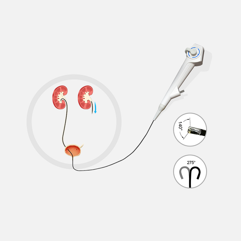
This electronic flexible ureteroscope is specially engineered for modern urological surgery. Adopting an ergonomic lightweight structure, it effectively reduces operator fatigue during long-duration procedures. Featuring a slim shaft and smooth tapered tip, the scope enables precise, atraumatic navigation inside narrow urinary anatomical passages
Equipped with an integrated video interface and high-clarity cold light transmission, the scope delivers bright, detailed visualization with no risk of thermal injury to surrounding tissues. It is well-suited for both diagnostic examinations and therapeutic ureteroscopic interventions.
With outstanding deflection performance, wide field of view and compact outer diameter, this electronic ureteroscope provides excellent operability and broad compatibility, serving as a reliable endoscopic device for routine urological clinical care.
Product Specifications
Outer diameter of main hose: ≤2.8mm
Inner diameter of clamp pipe: ≥3.0mm
Working length:680mm
Total length: ≥520mm
Field of view: 140° (direct view)
Observation depth:≥22-150mm
Resolution:≥520,000 pixels
Bending angle:≥270° upward (D) ≥ 180
Electronic Ureteroscope Advantage
This electronic ureteroscope features an ultra-slim shaft of ≤2.8mm outer diameter. The smooth tapered tip facilitates gentle navigation inside narrow urinary tracts and reduces trauma to patients. It boasts outstanding deflection capacity with upward bending up to 270° and downward bending 180°, allowing comprehensive observation across all renal calyces.
Equipped with a ≥520,000-pixel imaging sensor, it provides high-clarity real-time endoscopic images to assist clinicians in lesion identification. The ergonomic lightweight handle effectively alleviates operator fatigue during prolonged procedures. With integrated cold light transmission, there is no risk of thermal injury to tissue. The ≥3.0mm working channel supports matching laser fibers, biopsy instruments and other tools, meeting demands for diagnostic examination and minimally invasive urological treatment.
Electronic Ureteroscope Key Features
Ultra-slim Flexible Shaft
Outer diameter ≤2.8 mm with smooth tapered tip, enabling atraumatic access to narrow urinary anatomical structures.
Superior Bending Mobility
Upward bending ≥270°, downward bending ≥180°, realizing full observation of renal calyces.
High-resolution Imaging
Built-in ≥520,000 pixels sensor, providing sharp real-time endoscopic images for accurate lesion diagnosis.
Large Working Channel
Inner working channel ≥3.0 mm, compatible with laser fibers, biopsy forceps and other endoscopic accessories.
Wide Field of View
140° direct field of view, observation depth ranges from 2 mm to 150 mm for comprehensive examination.
Ergonomic Lightweight Handle
Reduces operator fatigue during long-duration urological surgeries and improves operation stability.
Safe Cold Light Visualization
Integrated video interface and cold light transmission avoid thermal tissue damage.
Complete Clinical Compatibility
Suitable for diagnostic ureteroscopy and minimally invasive therapeutic interventions in urology.
Electronic Ureteroscope Applications
Diagnostic Uses
Lesion Visualization
Biopsy Sampling
Therapeutic Uses
Stent Placement Assistance
Stone Fragmentation & Removal