


Views: 0 Author: Site Editor Publish Time: 2026-07-11 Origin: Site


Hospital procurement teams and clinical directors face mounting pressure to maximize capital equipment utilization. They must achieve this without compromising specialty-specific clinical outcomes. Purchasing siloed visualization equipment for every department creates redundant costs and complex maintenance protocols. It also causes workflow bottlenecks across ENT, orthopedics, general surgery, and gynecology. A strategically selected rigid endoscope can bridge multiple departments successfully. It must meet strict criteria for optical clarity, universal compatibility, and robust sterilization tolerance.
This article details the objective evaluation criteria required to select versatile endoscopy equipment. You will learn how to standardize optical specifications and ensure seamless camera integration. We also cover strategies to protect shared equipment from rapid degradation. Evaluating these elements carefully ensures you can support cross-disciplinary surgical workflows efficiently. By focusing on these parameters, facilities can streamline their capital investments. You will ultimately improve asset turnover while delivering exceptional patient care.
Multi-department suitability hinges on standardizing specific shaft diameters (e.g., 4mm and 10mm) and viewing angles (0° and 30°) that cover the majority of general and specialty procedures.
True cross-departmental efficiency requires seamless integration with an existing rigid endoscope camera system via universal couplers and standardized light post adapters.
Autoclavability and resistance to moisture ingress are critical; multi-department scopes experience higher reprocessing volumes, increasing the risk of rod-lens degradation.
Consolidating your purchasing volume fundamentally shifts how a hospital manages its surgical assets. Capital efficiency improves when facilities stop buying redundant imaging towers for every specialty. Standardizing equipment reduces your initial capital expenditure. It also simplifies vendor negotiations. You gain purchasing leverage by committing to a larger volume of standardized optical tools rather than fragmented, specialty-specific orders.
Workflow predictability improves dramatically alongside capital efficiency. Standardized equipment lowers the learning curve for everyone involved in the surgical loop. Rotating operating room (OR) staff face less confusion when setting up cases. Central sterile processing department (SPD) technicians handle familiar equipment daily. They develop muscle memory for specific inspection and cleaning protocols. This familiarity reduces handling errors and accelerates turnover times between surgical cases.
Transitioning from low-utilization, department-specific scopes to high-utilization, shared scopes improves your overall return on asset investment. A laparoscope sitting idle in general surgery could simultaneously serve a busy gynecology suite. Tracking utilization metrics across departments reveals how often siloed equipment remains unused. Shared scopes solve this inefficiency. They stay in constant circulation, maximizing the value derived from their operational lifespan.
We must acknowledge the clinical compromise limits transparently. Standardization works beautifully for the vast majority of cases. Roughly 80% of routine procedures can share standard optics. However, hyper-specialized procedures still require dedicated instruments. Neuroendoscopy or ultra-fine pediatric urology demands highly specific tools. You should not force standard scopes into these narrow applications.
To build a successful standardization strategy, follow these foundational steps:
Audit historical procedural volumes across all surgical departments.
Identify overlapping requirements for shaft diameters and viewing angles.
Consult lead surgeons to define acceptable optical baseline specifications.
Establish a unified SPD protocol for the newly shared equipment fleet.
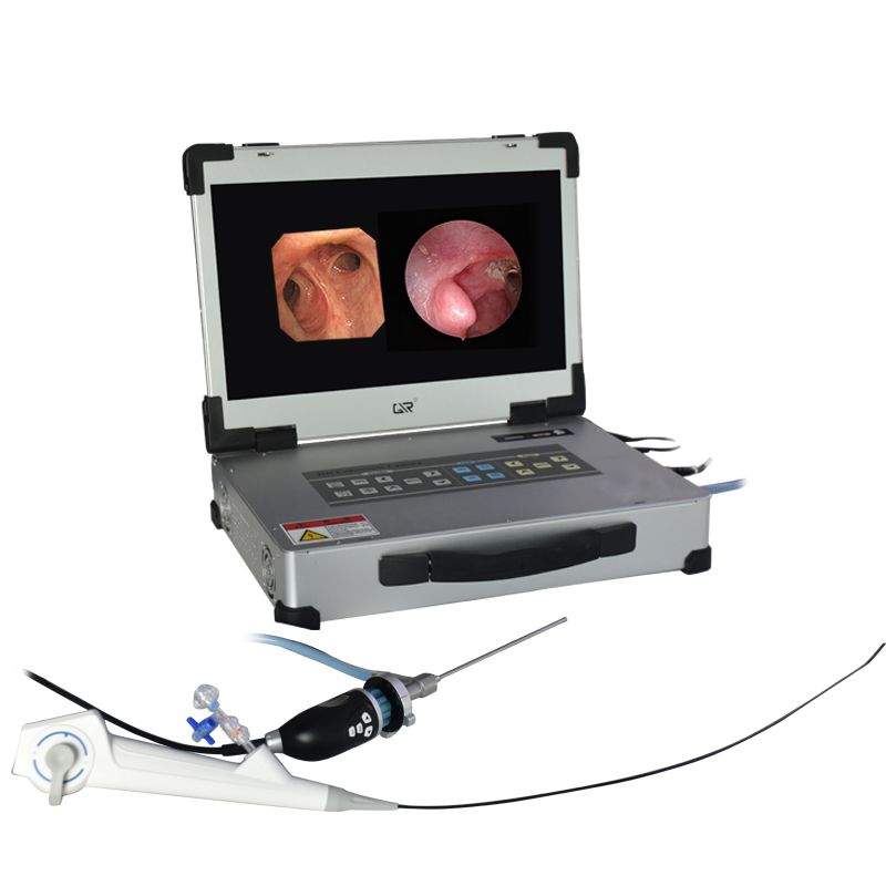
Selecting the right physical dimensions dictates how easily a scope transitions between specialties. Optimal shaft diameters serve as the foundation of multi-department utility. The 10mm scopes remain the gold standard for laparoscopy and general surgery. They maximize light transmission and provide a wide field of view. Surgeons rely on this robust diameter for complex abdominal and pelvic procedures where bright illumination is mandatory.
Conversely, 4mm scopes offer unparalleled versatility across smaller anatomical spaces. These thinner profiles dominate ENT for sinuscopy. They are equally vital in orthopedics for arthroscopy and in urology for cystoscopy. A facility bridging these diverse fields must invest heavily in high-quality 4mm units. They balance the need for minimal invasiveness with acceptable light delivery.
Direction of View (DOV) represents the second critical physical specification. Standardizing around two primary angles covers most clinical needs. The 0° lenses excel in forward-viewing procedures. Surgeons use them for standard laparoscopy and basic ENT diagnostic work. The straight-ahead view offers intuitive orientation. The 30° lenses provide an oblique visualization angle. They prove essential for navigating complex anatomical structures. Surgeons manipulate 30° scopes to look around corners during arthroscopy or complex abdominal procedures.
Underpinning these dimensions is the rod-lens architecture. Modern surgical interventions require high-fidelity, edge-to-edge resolution. The internal glass rod sequence must support varying depths of field across different surgical specialties. It must achieve this without peripheral distortion. A degraded peripheral image creates blind spots during delicate dissections. High-quality rod-lens designs maintain sharp focus whether the surgeon operates 2 centimeters or 10 centimeters away from the target tissue.
Standard Endoscope Diameters and Applications
Diameter | Primary Direction of View (DOV) | Target Clinical Specialties | Key Advantage |
|---|---|---|---|
10mm | 0°, 30°, 45° | General Surgery, Gynecology, Bariatrics | Maximum light transmission and field of view. |
5mm | 0°, 30° | Pediatric Surgery, Minimally Invasive Laparoscopy | Reduces port-site trauma while maintaining strong optics. |
4mm | 0°, 30°, 70° | ENT, Orthopedics, Urology | High versatility for navigating tight joint spaces and sinuses. |
2.7mm - 3mm | 0°, 30° | Small Joint Arthroscopy, Veterinary Surgery | Ultra-minimally invasive access for delicate structures. |
True cross-departmental efficiency relies on universal compatibility. You cannot achieve cost savings if new scopes require proprietary imaging towers. The scopes must feature standard ocular funnels. These universal eyecups connect seamlessly via C-mount couplers to any rigid endoscope camera system. A standardized DIN eyepiece ensures the optics lock securely onto the camera head. This tight mechanical seal prevents moisture from clouding the imaging sensor during a procedure.
Light source interoperability represents another common bottleneck. Purchasing versatile scopes means evaluating their light post adapters meticulously. Most high-end units include universal adapters compatible with Storz, Wolf, and ACMI fittings. This flexibility ensures the scope can accept existing fiber optic light cables across different OR suites. A scope moving from a urology suite to an ENT clinic must adapt instantly to whatever light source sits on the local equipment rack.
Sensor synergy dictates the final image quality displayed on the surgical monitor. The optical quality of the rigid optics must match the resolution capabilities of your existing camera control units (CCUs). Pairing a standard-definition legacy scope with a native 4K camera head creates a bottleneck. The high-resolution sensor only magnifies the optical flaws of the inferior lens. Conversely, premium 4K-rated optics ensure you extract maximum performance from advanced imaging processors.
Consider these best practices for seamless camera integration:
Verify that the C-mount coupler mechanism engages smoothly without grinding against the scope's ocular funnel.
Test light post adapters for secure threading to prevent light cable disconnections during active surgery.
Ensure the optical focal length matches the camera sensor size to prevent vignetting (dark corners on the monitor).
Standardize the anti-fogging protocols, as mismatched camera and scope temperatures often cause sudden condensation.
Multi-department scopes face a harsh reprocessing reality. Because they serve multiple disciplines, they undergo significantly more cleaning and sterilization cycles per week than dedicated specialty instruments. This high turnover rate exposes the equipment to accelerated thermal and mechanical wear. You must factor this relentless operational tempo into your procurement criteria.
Autoclave viability is non-negotiable for high-turnover scopes. Traditional chemical soaking takes too long for a scope shared across five daily cases. Facilities must select units specifically engineered to withstand repetitive high-temperature, high-pressure steam sterilization. Look for construction featuring laser-welded joints and advanced sapphire distal lenses. Laser welding eliminates the fragile adhesive seals prone to failing under steam pressure. Sapphire lenses resist the micro-scratching that degrades image clarity over hundreds of wash cycles. These features prevent moisture ingress, which remains the leading cause of optical failure.
Mechanical vulnerabilities multiply when scopes travel frequently. The primary risks include rod-lens cracking from bending stresses and severe distal tip damage. Surgeons occasionally use scopes as retractors in difficult cases, applying lateral force. This force shatters the delicate glass rods inside the shaft. Dropping the instrument or bumping the distal tip against a metal tray in the SPD also causes catastrophic damage.
Mitigation strategies must center on the sterile processing department. Establish rigorous inspection protocols to catch degradation early. SPD technicians should utilize dedicated endoscope testers. These simple devices verify light fiber transmission and check lens integrity before the scope reaches the autoclave. Catching a compromised seal before sterilization prevents pressurized steam from violently destroying the internal optics.
Lifecycle Risk vs. Mitigation Profile
Common Risk Factor | Primary Cause | Clinical Impact | SPD Mitigation Strategy |
|---|---|---|---|
Moisture Ingress | Seal failure during autoclave cycles. | Foggy or completely blurred surgical image. | Mandate leak testing before every wash cycle. Select laser-welded scopes. |
Rod-Lens Fractures | Lateral bending force or blunt impact. | Crescent-shaped shadows or total blackout in view. | Use rigid sterilization trays. Enforce strictly hands-off transport rules. |
Light Fiber Burnout | Excessive heat from light cables; physical crushing. | Dim image requiring dangerous light source increases. | Use light transmission testers. Avoid tight coiling of attached cables. |
Distal Tip Scratches | Contact with sharp surgical instruments or wire brushes. | Persistent glare or distorted peripheral tissue views. | Specify sapphire distal lenses. Restrict abrasive cleaning tools. |
Conducting a thorough needs assessment anchors the procurement process. You must audit procedural volume across laparoscopy, arthroscopy, and ENT divisions. Identify precise overlaps in required diameters and angles. Data drives these decisions. If your facility performs 400 arthroscopies and 300 sinus surgeries annually, prioritizing a robust fleet of 4mm 30° scopes makes perfect clinical sense.
Next, evaluate the vendor ecosystem versus an open architecture approach. Some manufacturers push a closed-loop proprietary ecosystem. In these setups, their scopes only function optimally with their proprietary camera heads and light cables. While this ensures out-of-the-box synergy, it restricts future flexibility. An open-architecture approach allows you to mix universal scopes with a surgical endoscope camera from various vendors. This flexibility protects your hospital from being locked into restrictive pricing models down the road.
You must scrutinize the warranty and repair infrastructure. High-use shared equipment will inevitably require maintenance. Assess the vendor's repair turnaround times carefully. Ask about their loaner equipment policies. If a shared 10mm scope goes down, you need a replacement within 24 hours to prevent canceling surgeries. Warranty coverage for fluid invasion is particularly critical. Review the fine print to ensure the manufacturer stands behind their autoclave compatibility claims.
Finally, mandate rigorous pilot testing. Never purchase shared optical equipment based solely on a brochure. Set up a cross-departmental trial spanning at least two weeks. Gather unbiased feedback from lead surgeons in different specialties. Ask them to grade the equipment specifically on ergonomic handling, center-to-edge illumination, and accurate color reproduction. Tissue color accuracy is vital for identifying ischemic bowel in general surgery or subtle mucosal inflammation in ENT. A scope that passes the rigorous standards of multiple specialists earns its place in the shared fleet.
Sourcing a rigid endoscope for multi-department use requires balancing physical dimensions, broad compatibility, and structural durability. You must prioritize features like sapphire lenses and standard C-mount compatibility. These elements ensure the equipment survives the demanding operational tempo of a shared asset pool.
Standardization proves highly cost-effective and improves workflow predictability across the hospital. This strategy succeeds perfectly provided procurement does not compromise the core optical integrity required by the most demanding specialty sharing the equipment. High-fidelity rod-lens architecture remains the non-negotiable foundation of this approach.
As a next step, we recommend auditing your current OR equipment compatibility. Map out your existing camera control units and light sources. Following this audit, schedule a multi-disciplinary clinical evaluation to test open-architecture scopes. Drafting a unified procurement specification sheet based on these trials will secure the best clinical and operational outcomes for your surgical teams.
A: While standard for laparoscopy, a 10mm scope is generally too large for most arthroscopic procedures, which typically require 4mm scopes. Multi-department setups usually standardize on a pairing of one 10mm and one 4mm system to safely cover both specialized disciplines.
A: Most rigid endoscopes use a standard DIN eyepiece that connects to universal C-mount couplers. However, always verify compatibility with your specific surgical endoscope camera to ensure a secure, moisture-tight fit and optimal focal length.
A: Increased utilization means more frequent handling and sterilization cycles, which can accelerate the wear of seals and rod-lenses. Robust repair agreements and strict SPD handling protocols are required to mitigate a shortened lifespan.
A: Most modern, high-quality rigid endoscopes are designed to be fully autoclavable. However, facilities must confirm the specific validated sterilization parameters (e.g., temperature and cycle time) provided by the manufacturer to maintain the warranty.


