
Language


Views: 0 Author: Site Editor Publish Time: 2026-07-13 Origin: Site


Surgical skill remains paramount in every operating room. However, clinical outcomes fundamentally rely on the visualization limits of your surgical equipment. Surgeons simply cannot treat what they cannot clearly see. Upgrading or standardizing surgical imaging often forces procurement teams to navigate highly fragmented specification sheets. You must frequently choose between proprietary vendor ecosystems and conflicting claims about optical clarity. Hardware longevity also complicates these decisions, making it difficult to find reliable benchmarks. This guide delivers an evidence-based, vendor-neutral framework for evaluating your next rigid endoscope system. We focus heavily on optical performance, component interoperability, and long-term equipment reliability. You will learn exactly how to align clinical needs seamlessly alongside hardware reality. We aim to equip you thoroughly for your next major equipment transition, ensuring every technical choice directly supports superior patient outcomes.
Selecting a rigid endoscope requires balancing optical resolution (field of view, depth of field) with mechanical durability (autoclave tolerance).
Seamless integration between the scope optics and the endoscope camera system is critical to minimizing image degradation.
True cost evaluation must extend beyond the initial purchase to include sterilization degradation rates, repair frequency, and cross-vendor compatibility.
Compliance, warranty terms, and validated reprocessing protocols act as primary risk-mitigation filters during the procurement phase.
Every surgical specialty demands unique optical performance metrics. You cannot apply a universal standard across completely different procedural environments. A laparoscopy requires a long working length and a wide field of view to navigate the abdomen safely. Conversely, arthroscopy and ENT procedures demand much shorter, narrower scopes to navigate tight joint spaces or sinus cavities. Mapping specific surgical requirements directly to hardware specifications ensures you select the correct viewing angles and shaft diameters from the start.
Many hospitals fall into the trap of upgrading their imaging sensors without updating their optical tubes. Purchasing a modern 4K endoscope camera system yields marginal clinical benefits if you pair it alongside legacy, standard-definition scopes. The physical rod-lens architecture inside older scopes acts as a severe bottleneck. It limits light transmission and spatial resolution. Ultimately, a 4K camera will simply capture a highly magnified, beautifully detailed image of a blurry optical output. You must upgrade both components simultaneously to unlock true high-definition visualization.
Establishing operational benchmarks early prevents long-term frustration. Clinical teams should outline baseline expectations for equipment uptime and sterilization turnaround times. Surgeon ergonomic fatigue also plays a critical role. Heavy camera heads or poorly balanced scopes lead to hand tremors during long operations. You should define success not just by image sharpness, but by how seamlessly the hardware supports the surgeon throughout a rigorous daily schedule.
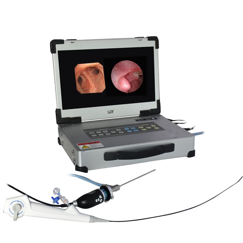
Understanding the internal architecture of a rigid scope empowers you to make better technical decisions. The optical train dictates image quality. Most modern designs utilize the Hopkins rod-lens system. Unlike conventional designs utilizing tiny lenses separated by large air gaps, the Hopkins system uses long glass rods separated by very small air spaces. This elegant inversion dramatically increases light transmission. It also reduces peripheral distortion, giving surgeons a clear edge-to-edge view.
Illumination mechanics represent another critical failure point. Fiber-optic light guide bundles travel along the outer edge of the scope shaft. They interface directly with external LED or Xenon light sources. Over time, thermal stress and physical bending cause individual glass fibers to snap. As more fibers break, light transmission drops significantly. This creates dark shadows around the edges of the surgical field. Evaluating the durability of these fiber bundles remains essential for maintaining bright, consistent illumination.
The camera coupling serves as the vital junction where the optics meet the sensor. You typically choose between universal C-mount couplers and proprietary locking mechanisms. Universal couplers offer immense flexibility. They allow you to attach scopes from different manufacturers to your existing camera heads. Proprietary locking systems restrict you to a single vendor. However, they often provide superior moisture seals and perfectly aligned optical centering. You must carefully weigh interoperability against the guaranteed optimization of a closed ecosystem.
Coupling Type |
Primary Advantage |
Primary Disadvantage |
Best Use Case |
|---|---|---|---|
Universal C-Mount |
Broad compatibility across various equipment brands. |
Potential for misalignment and higher moisture intrusion risk. |
Facilities maintaining diverse, multi-vendor equipment fleets. |
Proprietary Lock |
Optimal optical alignment and superior ergonomic balance. |
Locks the facility into a single manufacturer's ecosystem. |
Departments standardizing entirely on one premium brand. |
You cannot effectively evaluate optical fidelity on a sterile benchtop. You must assess center-to-edge sharpness and color fidelity under clinically relevant lighting conditions. In the operating room, deep cavities absorb light. Tissues reflect glare differently depending on moisture levels. A high-quality rigid endoscope system maintains crisp focus across the entire monitor, eliminating the "fish-eye" blurring common in cheaper models. Accurate red-color reproduction is especially vital for identifying subtle vascular structures.
Surgeons rely on specific viewing directions to safely navigate complex anatomy. Each angle presents distinct advantages and specific blind spots:
0° Scopes: Provide a straightforward, intuitive view. Best for forward navigation, but they cannot look around anatomical corners.
30° Scopes: Offer an oblique view. Rotating the scope expands the lateral field, making them the standard choice for general laparoscopy.
45° Scopes: Strike a balance for specialized joint navigation, frequently used in arthroscopy to view behind meniscal tissue.
70° Scopes: Deliver extreme lateral visualization. Essential for ENT sinus procedures, though they require high spatial awareness to avoid disorientation.
The physical environment inside a Central Sterile Services Department (CSSD) destroys fragile equipment. You must analyze the exterior materials rigorously. Premium scopes utilize laser-welded stainless steel shafts. They also feature scratch-resistant sapphire glass at the distal tip. Sapphire glass resists the abrasive wiping motions occurring during live surgery and manual pre-cleaning.
Demand validated data on autoclave endurance from your manufacturer. Repeated high-temperature steam sterilization inherently degrades internal optical adhesives over time. Thermal expansion forces the metal shaft and glass lenses to expand at different rates. This stress eventually breaks internal moisture seals. Once a seal fails, steam enters the optical train, causing permanent interior fogging. Selecting equipment engineered specifically to withstand thousands of harsh sterilization cycles drastically improves operational longevity.
The debate between open architecture and closed ecosystems heavily influences long-term facility flexibility. A single-vendor closed system guarantees that the light source, scope, and camera communicate flawlessly. Vendors can optimize color algorithms perfectly when they control every hardware piece. They also provide a single point of accountability when failures occur. You never have to deal with two different manufacturers blaming each other for poor image quality.
Conversely, open-architecture models offer significant agility. They provide the flexibility to use existing light cables and camera heads. If a specific department prefers a niche scope brand for a highly specialized procedure, an open system accommodates it smoothly. Facilities must decide whether they value seamless, locked-in optimization over the freedom to mix and match modular components.
Compliance acts as your strongest shield against operational risk. You must verify that any new endoscope camera system perfectly aligns with local regulatory standards, such as FDA clearances or CE markings. More importantly, the manufacturer's validated Instructions for Use (IFU) must align with your internal hospital CSSD protocols. If a vendor requires a sterilization chemical your hospital does not stock, implementation will stall immediately.
Physical handling vulnerabilities cause the majority of hardware failures. Rigid endoscopes are remarkably fragile instruments. Bending the shaft even a few millimeters shatters the internal glass rod-lenses instantly. Implementation strategies must address human error directly. Best practices include:
Deploying dedicated, custom-molded sterilization trays that lock scopes securely in place.
Mandating that heavy surgical instruments never share a sink basin alongside delicate optics.
Implementing regular staff training programs focused purely on safe transport and manual cleaning techniques.
Finally, modern surgical imaging relies heavily on digital infrastructure. Ensure the processing unit complies with rigorous data security standards like HIPAA or GDPR. The software must integrate smoothly with existing hospital PACS and EMR networks. Scrutinize the contract for hidden software licensing fees related to image storage or network connectivity. Secure, seamless image routing directly impacts post-operative documentation efficiency.
Framing the initial acquisition phase against ongoing maintenance realities reveals the true operational footprint of surgical equipment. Initial setups represent only a fraction of the overall lifecycle. You must focus intensely on expected repair frequencies and the overall lifespan of the optics. A system that looks spectacular on day one but spends weeks in the repair depot disrupts surgical schedules and increases departmental stress.
Evaluate vendor repair programs meticulously. Look for robust exchange programs and immediate loaner equipment availability. While third-party repair facilities sometimes offer faster turnarounds, Original Equipment Manufacturer (OEM) repairs generally preserve the original optical specifications. Third-party vendors may replace broken Hopkins rod-lenses with inferior conventional lenses, permanently downgrading the image quality without your knowledge. Prioritize service agreements that guarantee exact specification matching.
To navigate the procurement phase efficiently, adopt a pragmatic, three-step shortlisting framework:
Clinical Specification Matching: Cross-reference the technical capabilities (viewing angles, resolution, illumination) directly with the specific procedural demands of your surgical heads.
CSSD Workflow Validation: Ensure the required cleaning parameters and sterilization temperatures match your existing hospital infrastructure perfectly.
Lifecycle Stability Calculation: Review historical hospital breakage rates for similar equipment. Use this data to project the expected operational lifespan and determine the necessary volume of backup inventory.
The optimal rigid endoscope system consistently balances elite optical clarity with rugged, real-world reprocessing endurance. You cannot sacrifice mechanical durability for high resolution, nor can you accept subpar imaging simply to gain a stronger steel shaft. Success requires integrating superior rod-lens architecture seamlessly with highly advanced camera sensors. When these elements align, surgeons gain the precise visualization they need to improve patient outcomes safely.
Moving forward, procurement teams and surgical heads should always request in-vivo clinical trials. Never rely solely on perfectly curated technical specification sheets. Seeing how the equipment performs inside an actual operating room reveals nuances benchtop tests hide. Furthermore, encourage strict cross-departmental alignment. Surgeons, Biomedical Engineering, and CSSD staff must all agree on the hardware's usability and maintainability before drafting the final Request for Proposal (RFP). Collaborative evaluation ensures your chosen system performs reliably for years.
A: A high-quality rigid endoscope typically survives between 500 and 1000 autoclave cycles before noticeable optical degradation occurs. This range depends heavily on Original Equipment Manufacturer (OEM) build quality. During sterilization, extreme heat causes the metal shaft and interior glass components to expand at different rates. This continuous thermal expansion repeatedly stresses the optical adhesives. Eventually, these microscopic seals micro-fracture, allowing steam to penetrate the optical train and cause permanent internal fogging.
A: Yes, you can physically connect them using universal C-mount couplers. However, the physical rod-lens inside older scopes severely limits the actual resolution. Older scopes cannot transmit the massive amount of light and detail required for true 4K imaging. Consequently, the advanced camera sensor will simply capture a highly detailed, magnified image of a fundamentally lower-resolution optical output. Upgrading the optics is necessary for true 4K clarity.
A: A direct-coupled rigid endoscope features an integrated, non-removable camera head built directly into the scope shaft. This design eliminates the physical junction between components, significantly reducing internal fogging risks and improving ergonomic balance. However, if the optics break, the entire unit requires servicing. A modular system separates the scope and camera via a coupler. This allows higher repairability and flexibility, as you can easily swap out damaged scopes without replacing the expensive camera head.
A: You should prioritize comprehensive coverage for fluid invasion and thermal damage. These two factors represent the most common points of operational failure. Fluid invasion ruins the internal optics, while thermal damage often degrades fiber-optic light bundles and internal adhesives. Ensuring your warranty explicitly covers failures induced by standard, validated hospital sterilization cycles protects your facility against premature equipment degradation and unexpected downtime.


