| Availability: | |
|---|---|
| Quantity: | |
HD-390
Geiwre
The HD-390 All-in-One 4K Endoscopic Camera System is engineered to redefine surgical visualization by integrating cutting-edge 4K Ultra-High Definition technology into a highly compact, portable unit. This state-of-the-art system seamlessly combines a high-resolution 4K camera head, a powerful 140W LED light source, and an advanced image processing unit within a single, streamlined console. By eliminating the clutter of multiple separate devices and complex cabling, the HD-390 delivers unparalleled image clarity, stunning color accuracy, and exceptional depth of field directly to compatible 4K monitors.
Designed for maximum versatility and operational efficiency, it supports a wide range of rigid endoscopes (such as laparoscopes and arthroscopes). It serves as the ideal solution for modern operating rooms and clinics seeking premium endoscopic imaging in a user-friendly, space-saving package.
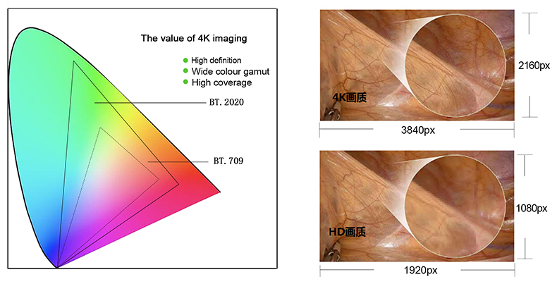
Based on the "Ultra High Definition (UHD)" standard defined by the International Telecommunication Union (ITU), the system features a physical resolution of 3840x2160 (4Kx2K). This provides twice the width and height, and four times the overall pixel area of standard Full HD (1920x1080). The resulting 4K resolution ensures exceptional detail capture during complex procedures.
Furthermore, the system adopts the BT.2020 color standard, which offers a significantly wider color gamut compared to conventional displays. For clinical applications, this enhanced color space allows for better discrimination of critical tissue boundaries, such as fat, nerve, and vascular junctions, enabling surgeons to clearly observe micro-vessels and pathological tissue contours.
| Camera | 9,000,000 Pixels 1*1.8 "CMOS |
| Resolution | 3840(H) *2160(V) |
Definition | 1300 Lines |
| Minimal illumination | 0.3Lux |
Video input | HDMI |
Video output | HDMI,DVI,3G-SDI,VGA,USB |
Shutter speed | 1/60~1/60000(NTSC),1/50~50000(PAL) |
Camera cable | 2.9m/Special lengths can be customized |
LED Light source | 140W |
| Long life | ≥50000Hours |
Power supply | 85 ~ 264VAC |
Monitor | 24''monitor |
Language | Chinese, English, Russian, French, Spanish, Portuguese can be switched |
Ultra-High Definition 4K Imaging: Delivers 3840x2160 resolution for exceptional clarity, precision, and depth of field, ensuring superior surgical visualization.
All-in-One Compact Design: Integrates a 9-megapixel 4K camera head, a 140W LED light source, and an image processor into a single, portable unit, significantly reducing OR clutter.
Stunning Color Accuracy: Utilizes the BT.2020 color standard to ensure true-to-life colors for accurate tissue and vascular differentiation.
Broad Endoscope Compatibility: Engineered to support a wide range of rigid endoscopes (laparoscopes, arthroscopes, etc.) for versatile clinical applications.
Space-Saving & Portable: Streamlined physical design ideal for operating rooms and outpatient clinics with limited space.
Seamless 4K Monitor Integration: Directly outputs high-quality, uncompressed imaging to compatible 24-inch 4K displays for real-time viewing.
HD-390 All-in-One Endoscopic Camera System: Workflow & Functional Advantages
The HD-390 All-in-One Endoscopic Camera System is engineered to streamline medical workflows by delivering instantaneous, crystal-clear visualization of internal anatomical structures. By seamlessly connecting with compatible endoscopic instruments, the system captures real-time visual data from the surgical area and projects it directly onto the integrated 24-inch monitor. This immediate, high-fidelity visual feedback is crucial for medical professionals, allowing them to observe subtle tissue variations and make accurate diagnostic decisions without delay. The integrated design not only reduces equipment clutter in the operating room but also minimizes setup time, ensuring that the surgical team can focus entirely on patient care and procedural safety.
To support complex procedures that demand meticulous attention to detail, this camera system features sophisticated image magnification and enhancement capabilities. Medical practitioners can easily enlarge specific areas of interest, allowing for a more detailed examination of micro-tissue structures and subtle lesion margins. Furthermore, the system automatically optimizes contrast, brightness, and color balance in real time. This dynamic optimization ensures that critical anatomical boundaries—such as fine blood vessels and nerve networks—remain highly distinguishable even under challenging lighting conditions within the body cavity. By providing a clearer and more differentiated view, the system significantly reduces visual fatigue for the surgeon and enhances overall procedural precision.
Successful surgical interventions rely heavily on the coordinated efforts of the entire medical team. The HD-390 system facilitates this by transmitting live visual feeds to the display monitor with virtually zero latency. This ensures that the primary surgeon, assisting physicians, and nursing staff all share the exact same visual perspective simultaneously. Such synchronized viewing is essential for anticipating the surgeon's next moves, preparing the correct instruments promptly, and maintaining smooth communication throughout the procedure. By eliminating visual discrepancies among team members, the system fosters a highly collaborative operating environment that contributes to improved surgical efficiency and reduced operation times.
Beyond real-time visualization, the system offers robust video recording and storage functionalities, adding significant long-term value to medical institutions. Entire examination processes or surgical procedures can be captured and securely saved for future reference. This built-in recording capability is an invaluable asset for medical education, allowing experienced surgeons to share practical case studies and techniques with medical students. Additionally, the stored footage serves as a reliable resource for post-operative case analysis, academic research, and maintaining accurate medical records. Having direct access to clear procedural recordings helps institutions ensure compliance, facilitate peer reviews, and provide objective documentation for medical dispute resolution.
Recognizing the diverse technological ecosystems present in modern healthcare facilities, the system is designed with extensive connectivity options. It supports a comprehensive range of multi-mode video outputs, including HDMI, DVI, SDI, and CVBS formats. This broad compatibility ensures that the camera system can be effortlessly integrated with various existing medical devices, secondary external monitors, surgical broadcasting networks, or hospital information systems. Procurement teams can be confident that the system will adapt to different operating room setups without the need for complex, third-party signal converters, thereby protecting existing equipment investments and ensuring flexible deployment across different clinical scenarios.
The HD-390 All-in-One Laparoscopic Endoscope Camera System is designed to meet the diverse and demanding requirements of modern minimally invasive surgery. By providing a highly detailed surgical field of view, this integrated system supports medical professionals across multiple clinical departments, ensuring precise diagnostics and effective therapeutic procedures. Below are the primary clinical applications where the system delivers significant value.
Thoracoscopy: Widely utilized for the diagnosis and treatment of various thoracic conditions. It plays a crucial role in the etiology diagnosis of pleural effusion, accurate staging of lung cancer, and the evaluation of pleural diseases. During complex thoracoscopic surgeries, the integrated endoscopic camera system provides thoracic surgeons with a highly defined surgical field. This visual clarity is essential to assist in delicate procedures such as lobectomy and mediastinal tumor resection, helping to identify critical anatomical structures and ensure safe tissue removal.
Cystoscopy: Serves as an important method to diagnose and manage bladder diseases, including cystitis, bladder stones, and bladder tumors. Through the cystoscope, doctors can closely observe the internal condition of the bladder mucosa. Furthermore, the system supports cystoscopic treatments, enabling precise interventions like bladder stone lithotripsy and transurethral resection of bladder tumors.
Ureteroscopy: Applied for the diagnosis and treatment of ureteral diseases, such as ureteral calculi and ureteral stricture. When performing lithotripsy or stricture dilation under a ureteroscope, the camera system helps doctors operate accurately within narrow anatomical spaces. This visual support is vital for improving the overall success rate of the operation and reducing the risk of postoperative complications.
Hysteroscopy: Essential for diagnosing and treating lesions within the uterine cavity, such as endometrial polyps, uterine fibroids, uterine adhesions, and uterine malformations. In hysteroscopic surgery, the doctor can perform precise manipulation based on the highly detailed image displayed on the monitor. This allows for the targeted removal of diseased tissue while carefully preserving the normal physiological function of the uterus.
Laparoscopy: Widely used in the comprehensive diagnosis and treatment of gynecological diseases, including ovarian cysts, uterine fibroids, and endometriosis. Laparoscopic surgery offers the clinical advantages of less surgical trauma and quicker patient recovery. The integrated endoscopic camera system ensures a clear, unobstructed view of the pelvic cavity, which is necessary to maintain the safety and accuracy of the operation during complex tissue dissection.
Nasal Endoscopy: Enables specialists to clearly observe the complex structures and potential lesions of the nasal cavity and sinuses. It is highly effective for identifying sinusitis, nasal polyps, and nasal tumors, providing a reliable basis for disease diagnosis and treatment planning. In nasal endoscopic surgery, the system helps doctors accurately remove diseased tissue while retaining the normal nasal structure and its essential functions.
Laryngoscope: Used to carefully examine lesions of the larynx, such as vocal cord polyps, vocal cord nodules, and early-stage laryngeal cancer. For certain laryngeal conditions, minimally invasive surgeries can be directly performed under the laryngoscope, such as vocal cord polypectomy, where precise visualization is required to protect vocal function.
Arthroscopy: Widely applied in the diagnosis and surgical treatment of joint diseases affecting the knee joint, shoulder joint, and hip joint. Common indications include articular cartilage injury, meniscus injury, and ligament tears. Arthroscopic surgery involves the insertion of arthroscopes and specialized surgical instruments through small incisions. With the aid of the camera system, the orthopedic surgeon can clearly visualize the internal structure of the joint on the monitor, facilitating accurate repair and complex reconstruction surgeries with minimal disruption to surrounding tissues.
Do I need a specific 4K monitor to fully utilize the HD-390 system?
Yes. To experience the true clinical benefits of the 4K UHD resolution, the system must be paired with a compatible 4K monitor that supports the required input types (such as HDMI 2.0) and matches the resolution and frame rate output of the HD-390. While the system is backward compatible and will function with standard HD monitors, the visual output will be limited to standard HD resolution in those setups.
Is the camera head compatible with standard sterilization processes like autoclaving?
The camera head is specifically engineered to withstand rigorous medical sterilization protocols. It is designed to be compatible with standard methods, including autoclave and hydrogen peroxide plasma sterilization. For optimal equipment longevity and patient safety, we strongly recommend verifying the specific temperature, pressure, and cycle requirements detailed in the manufacturer's Instructions for Use (IFU) prior to processing.
What types of rigid endoscopes are compatible with this camera system?
The HD-390 is designed for broad compatibility across various surgical disciplines. It works effectively with most standard rigid endoscopes available on the market, utilizing universal interfaces such as the standard C-mount or specific bayonet mounts. This universal design helps integrate the system into your existing medical equipment inventory without requiring immediate scope replacements, reducing overall procurement costs.
How powerful is the integrated LED light source for deep cavity procedures?
The system is equipped with a high-intensity integrated LED light source capable of delivering illumination exceeding 180,000 lux at the scope tip. This exceptional brightness ensures clear, well-illuminated visualization even in deep anatomical cavities or during demanding laparoscopic procedures where adequate lighting is critical for surgical precision and safety.
Can the HD-390 system be used to record 4K surgical videos?
Yes, 4K video recording is fully supported. By connecting the system's high-definition video output interfaces (such as HDMI or SDI) to an external 4K-capable medical video recorder or capture device, surgical teams can easily capture and store high-resolution footage. This is highly beneficial for patient documentation, postoperative case reviews, and academic medical training purposes.