


Views: 0 Author: Site Editor Publish Time: 2026-07-22 Origin: Site


Visualization remains the undeniable cornerstone of successful urological endoscopy today. Surgeons rely entirely on pristine optics to navigate delicate urinary tracts and target stones safely. Without absolute visual clarity, even the most routine procedures become hazardous. Unfortunately, substandard optics and poor device design frequently cause extended operating room (OR) times. They also lead to incomplete stone clearance and significantly elevate the risk of ureteral trauma. When your tools fail to deliver a crisp image, patient safety hangs in the balance.
This article provides clinical directors and surgical teams with a comprehensive framework for evaluating essential equipment features. We will explore optical superiority, mechanical durability, and critical integration requirements. You will learn exactly how to evaluate a rigid ureteroscope to maximize procedural safety, boost efficiency, and ensure long-term reliability. By prioritizing these structural and optical elements, hospitals can streamline surgical workflows and vastly improve patient outcomes.
Optical superiority: Rod-lens systems provide the highest resolution for rigid devices, directly impacting procedural safety.
Design tradeoffs: Balancing working channel diameter with outer shaft size is critical for maintaining continuous irrigation without requiring ureteral dilation.
Vendor evaluation: Procurement decisions must weigh initial capital expenditure against repair service agreements and tower compatibility.
Urology departments measure procedural success through two distinct lenses. Clinical outcomes dictate patient safety, while operational efficiency drives department viability. You must balance both to run a successful endoscopic program.
Clinical Success Criteria: Achieving high stone-free rates (SFR) remains the primary clinical objective. Surgeons must fragment and extract stones completely. They must also minimize mucosal injury and prevent postoperative complications like strictures. An excellent visual field allows the surgeon to identify tiny stone fragments. It also helps them avoid accidental thermal damage during laser lithotripsy. When you can see clearly, you can operate safely.
Financial Success Criteria: Modern surgical centers face immense pressure to reduce per-procedure costs. Financial success relies heavily on efficient OR turnover. You must minimize equipment downtime and reduce repair frequencies. Reliable instruments keep the daily surgical schedule moving. Frequent equipment failures disrupt these schedules and create expensive delays. You can achieve strong financial performance simply by selecting highly durable instruments.
The Visualization Bottleneck: Poor image quality serves as the primary bottleneck in ureteroscopy. Optical degradation directly correlates with increased surgical risk. Cloudy lenses obscure the surgical field. Broken optical fibers create dark spots on the monitor. These visual impairments force surgeons to work slower. Prolonged procedures increase anesthesia times for the patient. They also increase the likelihood of fluid absorption complications. Eliminating this visualization bottleneck remains crucial for improving overall surgical metrics.
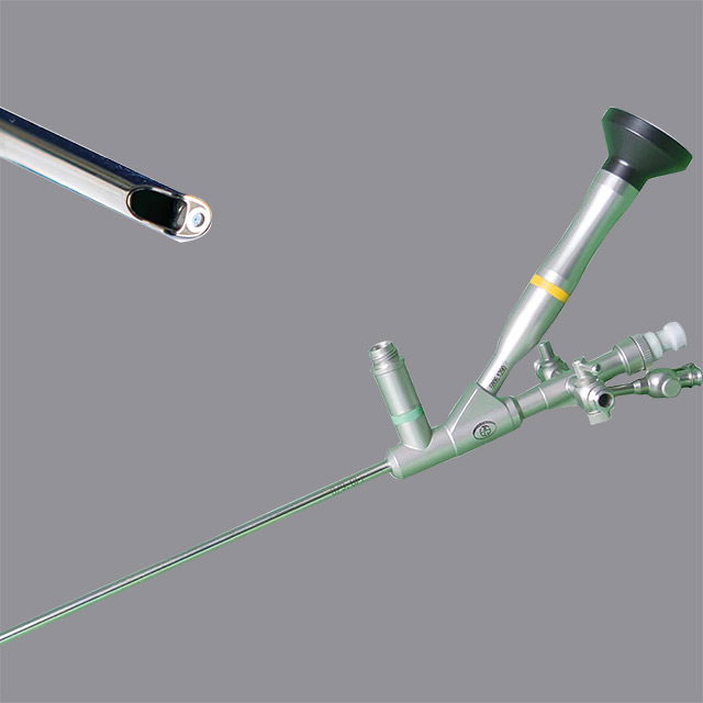
Optical quality dictates every movement a urologist makes inside the ureter. Traditional rod-lens technology remains the gold standard for high-definition imaging in rigid instruments. This system uses specialized glass rods separated by tiny air spaces. It delivers a bright, distortion-free image to the camera sensor. The resolution provided by a rod-lens system is unparalleled. It allows surgeons to distinguish subtle tissue variations and track fine laser fibers precisely.
Durability realities present a clear contrast between technologies. Fiber optic bundles are inherently susceptible to progressive breakage. Over time, individual fibers snap due to normal bending and handling. This creates permanent black spots on the surgical monitor. These spots degrade the image quality permanently. Conversely, rod lenses do not degrade slowly. However, they are highly vulnerable to physical shock. Dropping a rod-lens instrument usually shatters the internal glass entirely. Understanding these characteristics helps you choose the right tool for your specific handling environment.
Feature | Rod-Lens Technology | Fiber Optic Bundles |
|---|---|---|
Image Resolution | Exceptional clarity, distortion-free edges | Good, but often displays a visible pixel grid |
Light Transmission | Superior brightness in deep cavities | Adequate, diminishes as fibers break |
Failure Mode | Immediate failure upon severe impact (shattering) | Gradual degradation (black spots over time) |
Flexibility | Rigid only | Allows for semi-rigid bending |
You cannot treat what you cannot see. Evaluating light fiber distribution is vital for achieving shadow-free illumination. High-quality instruments distribute light-emitting fibers evenly around the objective lens. This concentric design eliminates dark zones in the operative field. It provides uniform brightness across the entire mucosal wall.
Compatibility with high-output light sources also demands attention. Modern operating rooms utilize powerful LED and Xenon light sources. The instrument's light guide must safely transmit this intense energy without overheating the distal tip. Poor light transmission causes thermal hazards inside the ureter. Always ensure your chosen instrument handles high-intensity illumination safely and efficiently.
Best Practice: Always inspect the light post connection before surgery. Burned or degraded light fibers at the post drastically reduce illumination inside the patient.
The structural tradeoff between working channel size and shaft diameter requires careful consideration. You need a large working channel to accommodate laser fibers, stone baskets, and guidewires. However, maximizing this channel often means increasing the outer diameter of the instrument. A larger outer shaft makes insertion much more difficult.
You must maintain sufficient space for continuous irrigation flow. Irrigation clears surgical debris, stone dust, and blood from the visual field. If an instrument occupies the entire working channel, irrigation stops. The visual field immediately clouds over. Opt for designs boasting high-flow channels. These specialized designs allow pressurized fluid to bypass inserted tools. This ensures you maintain a clear view during aggressive lithotripsy.
Distal tip design significantly influences the ease of patient access. Manufacturers often design tips to be beveled or bullet-shaped. A smooth, atraumatic profile facilitates easier entry into the ureteral orifice. It gently dilates the natural tissue without causing microscopic tears. This specific shape helps you navigate past tight strictures safely.
The clinical benefit of an atraumatic tip is substantial. It frequently reduces the need for routine ureteral dilation. It also minimizes the reliance on ureteral access sheaths in straightforward distal cases. By avoiding unnecessary dilation, you protect the patient from prolonged swelling and potential stricture formation later. Smooth navigation translates directly to faster, safer surgeries.
Urologists constantly navigate the tortuosity of the ureter. Deciding between semi-rigid and purely rigid profiles is a daily clinical choice. Purely rigid instruments offer superior control for pushing past large, impacted stones. However, they struggle to navigate sharp bends in the male anatomy. Semi-rigid profiles incorporate slight flexibility. This allows you to navigate pelvic brims without compromising the instrument's structural integrity.
Ocular ergonomics play a massive role in surgical performance. The eyepiece design impacts surgeon fatigue during prolonged, complex procedures. Angled eyepieces allow the surgeon to maintain a comfortable neck posture. Straight eyepieces often force operators into awkward positions. Reducing ergonomic strain helps surgeons maintain intense focus during critical moments of stone fragmentation.
Adding new instruments to an existing surgical fleet requires careful compatibility checks. You must assess how a new device integrates with your legacy urological endoscope ecosystems. Operating rooms already feature established camera heads, light cables, and video monitors. The new instrument must connect seamlessly to these existing components.
Avoiding forced ecosystem lock-in protects your department's budget. Some manufacturers design proprietary connections. These force you to buy their specific camera heads and light cables. Look for instruments featuring standard DIN or universal ocular connections. This standardization ensures you can mix and match equipment as technology evolves.
Central sterile services departments (CSSD) handle instrument reprocessing daily. You must evaluate the requirements for autoclavability versus chemical sterilization. Autoclavable instruments offer rapid turnaround times. They withstand high-pressure steam, getting them back to the OR quickly. However, repetitive high-temperature sterilization cycles carry hidden risks.
Over time, intense heat causes optical degradation. The thermal expansion and contraction stress the internal glass components. Additionally, repeated steam exposure can cause adhesive failures at the distal tip. Once the seals break, moisture enters the optical channel. This renders the instrument completely useless. Ensure the manufacturer guarantees durability across hundreds of autoclave cycles.
Mechanical damage outside the OR remains a massive problem. Most instrument failures occur during transportation and cleaning, not during surgery. You must recognize these common points of failure to protect your inventory.
Staff training remains the most effective mitigation strategy. Implement rigorous training protocols for handling these delicate devices.
Transport safely: Always transport instruments in dedicated, padded rigid containers. Never stack heavy trays on top of them.
Handle with two hands: Support the heavy ocular end and the delicate shaft simultaneously during transfer.
Avoid bending forces: Never rest tools on the thin shaft during manual brushing and cleaning in the sink.
Inspect pre-sterilization: Use a magnifying loupe to check the distal tip for micro-cracks before sealing the tray.
Surgical success depends entirely on anatomical targeting. Clear clinical guidelines dictate instrument selection based on stone location. You should utilize rigid or semi-rigid scopes primarily for distal and mid-ureteral stones. These locations offer a relatively straight pathway. The stiff shaft easily pushes past swollen mucosa to reach the target.
Conversely, you must reserve flexible scopes for the proximal ureter and the renal pelvis. The upper urinary tract contains complex calyces and sharp angles. A stiff instrument simply cannot navigate into the lower pole of the kidney. Attempting to force a stiff shaft into these areas causes severe trauma. Proper allocation ensures optimal patient safety.
Hospital administrators monitor the baseline reality of surgical equipment expenses carefully. Rigid instruments offer a significantly lower cost-per-use compared to their flexible counterparts. They feature robust metal construction and lack delicate articulation mechanisms. This makes them inherently tougher.
Because they are tougher, they suffer a much lower repair frequency. Flexible instruments break easily if the laser fires while the tip is deflected. Rigid instruments avoid this specific failure mode entirely. By maximizing the use of stiff scopes for lower stones, departments can drastically reduce their overall equipment maintenance burden.
Hospitals should balance their inventory based on historical case-mix data. You must analyze your department's procedural history. Do your surgeons perform more distal ureteroscopies or complex intrarenal surgeries?
If distal stones dominate your schedule, invest heavily in highly durable metal instruments. If you serve as a tertiary referral center for complex kidney stones, you need a larger fleet of flexible devices. Smart fleet management ensures you never delay a case due to a lack of appropriate equipment. It aligns your inventory directly with patient needs.
Procurement teams must look far beyond the initial purchase price. Evaluating vendor service level agreements (SLAs) determines the true value of the equipment. You must scrutinize the vendor's guaranteed repair turnaround times. A broken instrument sitting at a repair facility helps no one. Fast turnarounds keep your OR running smoothly.
Loaner equipment policies also require careful review. Premium vendors automatically overnight a loaner instrument while yours undergoes repair. This policy prevents costly OR cancellations. Furthermore, analyze the specific warranty inclusions. Ensure the warranty covers optical degradation and moisture invasion, not just manufacturing defects.
Subjective opinions often cloud procurement decisions. You must standardize the evaluation process to gather actionable data. Setting up a blind clinical trial in the OR provides the most accurate assessment. Mask the brand names if possible, and ask multiple surgeons to grade the devices.
Focus the evaluation on three distinct parameters. First, assess image sharpness at the edges of the monitor. Second, evaluate handling traits, specifically the balance and ergonomics of the eyepiece. Third, test the irrigation flow rate while a standard laser fiber occupies the working channel. This standardized feedback removes bias and identifies the best clinical tool.
Once you complete the clinical trials, narrow your choices to two final vendors. Request comprehensive lifecycle repair expectations from these shortlisted companies. Ask them to provide historical data on the average number of uses before a major repair is required.
Review their proposed service agreements over a targeted 5-year period. Cross-reference their expected maintenance timelines with your department's projected case volume. Armed with surgeon feedback and transparent maintenance expectations, you can make a highly informed, confident purchasing decision.
Successful urological endoscopy demands a relentless focus on visualization and reliability. Selecting the proper equipment dictates both the safety of your patients and the efficiency of your surgical schedule. When you evaluate these tools systematically, you eliminate the guesswork from procurement.
Prioritize optics: Superior rod-lens systems eliminate the visualization bottleneck, drastically reducing surgical risk and anesthesia time.
Demand smart design: Instruments must balance a spacious working channel with an atraumatic tip to ensure constant irrigation and easy patient access.
Protect your inventory: Implement rigorous staff training for reprocessing to prevent bent shafts and optical degradation.
Evaluate holistically: Base your final decisions on hands-on surgical feedback and robust vendor service agreements rather than capital price alone.
Take action today by standardizing your next OR trial. Gather your surgical team, apply this evaluation framework, and select an instrument that truly elevates your clinical capabilities.
A: A high-quality instrument typically lasts between 100 and 150 procedures before requiring significant maintenance. However, this lifespan depends entirely on handling and sterilization methods. Aggressive manual scrubbing or frequent high-temperature autoclaving accelerates optical degradation. Implementing strict handling protocols and utilizing low-temperature sterilization can extend this operational lifespan significantly.
A: A purely rigid scope uses a solid metal tube and fragile glass rod lenses. It cannot bend. A semi-rigid scope incorporates flexible fiber-optic imaging bundles encased within a stiff but slightly pliable metal sheath. This design allows the shaft to bend gently under pressure, helping surgeons navigate the natural curves of the pelvic brim safely.
A: Standard outer diameters typically range from 6.0 Fr to 9.5 Fr. The ultra-slim 6.0 Fr scopes easily access tight ureters without prior dilation, making them ideal for pediatric or narrow anatomy cases. The larger 8.0 Fr to 9.5 Fr scopes provide maximum working channels for larger laser fibers and high-flow irrigation during aggressive lithotripsy.
A: Most modern instruments feature a universal DIN standard ocular eyepiece. This allows standard, commercially available camera heads to attach seamlessly using a standard c-mount coupler. However, certain specialized camera ecosystems may require specific proprietary adapters. Always verify cross-brand compatibility with your current video tower before purchasing.


