


Views: 0 Author: Site Editor Publish Time: 2026-07-15 Origin: Site


Endoscopy suites face compounding daily pressures. Procedure volumes steadily climb across global healthcare systems. Strict quality benchmarks demand consistently high adenoma detection rates. Meanwhile, physicians battle increasing physical fatigue during long shifts. Upgrading equipment often introduces unexpected workflow friction. New features might require extensive operator training initially. They can disrupt established reprocessing protocols significantly. This creates immense hesitation among clinical directors. Selecting the right colonoscopy machine requires looking beyond isolated imaging specs. Real return on investment emerges differently. You must evaluate mechanical design elements thoroughly. We must understand how AI integration affects cognition. Digital interoperability shifts the entire procedural sequence. This article guides you through modern endoscopy equipment evaluation. You will learn to identify true operational bottlenecks. We will explore key technical advancements. Ultimately, you will discover ways to enhance diagnostic accuracy. You will also improve room turnover times simultaneously.
High-definition imaging alone has diminishing returns; the current standard of evaluation must include real-time AI assistance (CADe/CADx) and its false-positive rates.
Ergonomic advancements in the electronic colonoscope directly reduce physician fatigue and can shorten cecal intubation times.
Infection control features and reprocessing compatibility dictate room turnaround times, a primary bottleneck in high-volume clinics.
Balancing high daily throughput creates conflicts against rigorous mucosal inspection. Standard benchmarks require meticulous examination. Physicians cannot rush the withdrawal phase ever. You need sufficient time to spot flat lesions. Yet, waiting rooms remain full constantly. Administrators push for faster room turnover. This creates a stressful environment for everyone.
We must identify true bottlenecks to solve this. Some delays happen strictly during the procedure. Looping creates significant intra-procedural friction. Difficult intubations consume valuable schedule minutes. Poor bowel preparation obscures the visual field. Other delays remain purely operational. Complex equipment setup slows down nursing teams. Manual EHR data entry wastes physician time. Sterilization workflows frequently stall suite operations. Disentangling these delays helps isolate necessary hardware features.
How do you measure upgrade success? You need a measurable reduction regarding total room time per patient. This reduction cannot compromise polyp detection capabilities. Ideally, new equipment improves your baseline ADR. We evaluate success through workflow fluidity. We want smoother insertions and faster lesion characterization.
Table: Classification of Endoscopy Workflow Bottlenecks
Category | Specific Bottleneck | Workflow Impact |
|---|---|---|
Intra-procedural | Complex loop formation | Delays cecal intubation heavily |
Intra-procedural | Poor mucosal visibility | Increases withdrawal inspection time |
Operational | Manual data entry | Delays physician transition between rooms |
Operational | Complex reprocessing steps | Extends overall room turnover time |
Establish baseline metrics before testing new devices. You cannot measure improvement without knowing current withdrawal times. Track your room turnaround delays meticulously for two weeks. This data highlights your most urgent operational needs clearly.
Focusing purely on procedural speed remains dangerous. Rushing withdrawal inevitably lowers adenoma detection rates. You must balance mechanical efficiency against thorough visual inspection. Speed should come from better tools, not faster rushing.
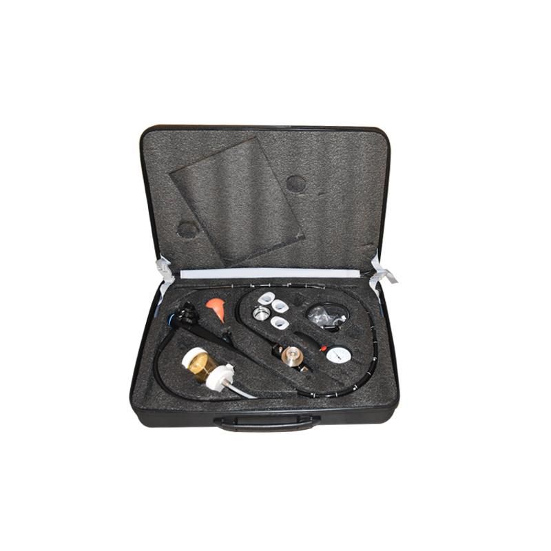
The shift to digital modalities changed everything recently. The modern electronic colonoscope outpaces legacy systems entirely. Older platforms coupled light sources directly via fiber optics. Modern designs decouple the illumination source smartly. They utilize advanced digital image processing instead. High-definition sensors sit directly at the distal tip. They transmit crisp video feeds instantly. This prevents image degradation during transmission. It allows for clearer, brighter displays.
Optical chromoendoscopy eliminates messy dye sprays. Narrow-band imaging illuminates specific vascular patterns clearly. Multi-LED technologies enhance mucosal surface contrast instantly. Physicians characterize tissues in real-time. This saves several minutes per polyp discovered. You no longer wait for dye diffusion. You can make immediate optical biopsy decisions.
AI-Assisted Detection acts as an expert second observer. Computer-aided detection (CADe) highlights suspicious areas visually. Computer-aided diagnosis (CADx) predicts histology instantly. These tools reduce cognitive load during withdrawal. Physicians experience less fatigue during afternoon lists. This sustains high detection rates all day long. They help equalize performance between junior and senior doctors.
You must evaluate AI algorithms carefully. Assess latency during rapid tube movements. False-positive rates matter deeply. Excessive false alerts cause severe alarm fatigue. They paradoxically increase total procedure time. The system flags normal folds continuously. Physicians must pause to verify each alert. This creates tremendous frustration and slows the schedule.
Implementation carries specific visual risks constantly. The AI requires seamless monitor overlay. It cannot distract the endoscopist's primary line of sight. Poorly designed interfaces clutter the main screen. They frustrate physicians rather than assist them. A clean UI guarantees better clinical adoption.
Variable stiffness technology revolutionizes insertion control. You can adjust the insertion tube’s flexibility on demand. This adjustment minimizes looping occurrences immediately. It reduces patient discomfort significantly. Efficient insertion directly impacts cecal intubation speed. Nurses spend less time applying manual abdominal pressure. The physician navigates complex sigmoid colons smoothly. This protects patient safety.
Wide-angle viewing expands visual boundaries massively. Modern lenses offer up to 170 degrees of vision. Retrograde viewing capabilities expose hidden areas easily. You can visualize blind spots behind mucosal folds. This requires far less excessive torque on the shaft. Physicians maintain better spatial orientation constantly. They catch hidden adenomas effortlessly.
Assess compatibility regarding physical distal attachments. Many physicians use cuff devices routinely now. These devices flatten folds during withdrawal effectively. They stabilize the scope tip during complex polypectomies. Your chosen system must accommodate these attachments securely. Incompatible tips render expensive accessories completely useless. Always verify attachment diameters before purchasing.
Handle ergonomics dictate physician career longevity. Poor weight distribution causes severe hand strain over time. Awkward dial placements force unnatural thumb stretches. High-volume endoscopists face severe repetitive strain injuries. Lighter handles mitigate these exact physical risks. Well-placed angulation controls reduce tendon stress daily. Physicians maintain finer control during delicate resections. This translates to safer patient outcomes.
Reprocessing dictates your daily patient volume capacity. Turnaround times create the primary suite bottleneck. Designing for decontamination requires strategic component choices. You must evaluate single-use components thoroughly. Disposable endcaps eliminate manual brushing needs completely. Sterile biopsy valves prevent cross-contamination inherently. Traditional reusable parts require intensive scrubbing. They delay room availability significantly. Transitioning to disposables speeds up bedside pre-cleaning.
Water jet systems maintain procedural momentum effortlessly. Dedicated forward water jets clear debris rapidly. You do not need accessory swaps to wash mucosa. A clear visual field prevents diagnostic delays. Efficient suction channels remove fluid without clogging. Physicians complete inspections without frustrating interruptions. This keeps the procedure moving forward.
Automated Endoscope Reprocessor compatibility remains critical. You must validate integration immediately. The new equipment must fit existing sterilization basins properly. Incompatible channel connectors prevent effective automated cleaning. This creates severe bottlenecks inside the sterile processing department. Your entire schedule stalls when scopes pile up.
We recommend evaluating the following compatibility points:
Verify connector block compatibility alongside existing basin dimensions.
Confirm channel pressure parameters match current pump outputs.
Check chemical compatibility regarding peracetic acid or glutaraldehyde.
Evaluate tracking software integration for infection control audits.
Image management software requires clean interoperability. Reporting systems must integrate deeply. They need to connect into existing EHR systems seamlessly. PACS integration ensures immediate image archiving. Manual image transfers waste precious turnaround minutes. Automated data flows reduce transcription errors significantly. They allow physicians to dictate reports immediately.
Legacy system compatibility protects previous capital investments. Upgrading from an older electrical colonoscope poses integration challenges. You must evaluate the required overhaul scope carefully. Does the transition require entirely new video processors? Must you replace all legacy light sources? Backward compatibility often exists within specific vendor ecosystems. This compatibility reduces financial shock significantly. You can phase your equipment upgrades gradually.
Service Level Agreements define your operational security. You must demand strict uptime guarantees from vendors. Ask about loaner scope availability during repairs. Determine the frequency of software updates. AI algorithms require continuous data refinement. Outdated software diminishes diagnostic advantages quickly. Solid vendor support keeps your suite running.
Procurement demands real-world clinical trials always. You cannot rely on showroom demonstrations alone. Showrooms lack the pressure of a packed waiting room. Follow this proven evaluation sequence:
Initiate clinical trials within your actual endoscopy suite.
Measure baseline metrics including ADR and withdrawal times.
Track average room turnaround times across one typical week.
Deploy the trial equipment under normal patient loads.
Compare trial data against baseline metrics before vendor selection.
Successful equipment evaluation requires a strategic mindset. You must prioritize features offering measurable workflow enhancements. Avoid chasing technical specifications without practical clinical value. AI-driven systems provide essential cognitive relief during long shifts. Mechanical efficiency speeds up intubation phases safely. Rapid turnaround designs keep schedules moving reliably. We encourage holistic evaluation processes always. Involve clinical endoscopists early in the discussion. Include sterile processing staff during hands-on trials. Holistic adoption prevents post-purchase workflow disruptions. Your next equipment upgrade should empower your entire clinical team.
A: It may slightly increase withdrawal time initially. This happens due to higher detection rates of diminutive polyps. However, it reduces time spent characterizing lesions. If you utilize CADx effectively, overall efficiency improves. Physicians spend less time guessing histology. The system creates a smoother, more confident workflow.
A: "Electrical" historically refers to older fiber-optic systems. These required external electrical light sources to illuminate tissue. "Electronic" refers to modern digital videoscopes. They feature CCD or CMOS chips embedded directly at the distal tip. This enables direct digital video transmission. It produces a much higher resolution image.
A: Yes, clinical evidence strongly supports this. Variable stiffness reduces the occurrence of complex loops. This directly decreases the time required to reach the cecum. It also reduces the need for manual abdominal pressure from nursing staff. The procedure becomes less physically demanding for everyone involved.
A: You should focus on automated image capture. Images must transfer directly into the patient's chart. Prioritize strict HL7 and DICOM compliance. This eliminates tedious manual data entry post-procedure. Seamless integration prevents transcription errors. It frees up staff to prepare the next patient faster.


